Emergancy Ortho: Elbow
The Elbow
ATRAUMATIC ELBOW PAIN:
Most often this can be diagnosed in 30 seconds with point tenderness and resistance of wrist flexion and extension. This is because the most common tendonitis or overuse injuries at the elbow are actually the insertion of the common flexor and common extensor tendons of the wrist and hand. Medial epicondylitis (i.e. Golfer's elbow) is tenderness at the medial epicondyle and pain there with resisted elbow flextion, while lateral epicondylitis (Tennis elbow) is pain at lateral epicondyle with palpation and resisted wrist extension. No x-rays are required and wrist splinting with pressure bands at the proximal tendon with ice and NSAIDs are the treatment of choice. Of course, the biceps and triceps tendons can have tendonitis and this is demonstrable on exam as well. There will be tenderness of these tendons and pain with resisted movement. The triceps is responsible for elbow extension and the biceps brachii can be isolated by having the patient flex the elbow and supinate against resistance (I grasp their hand like a handshake to add resistance). Sometimes olecranon bursitis results without trauma or very mild trauma and is again easy to see, palpate, and diagnose. Much consternation arises with aspiration of these bursa or not, so I will duck out of the debate and say that rarely would I aspirate because those whom I am concerned have a septic bursa look like there could be overlying cellulitis and I don't want to seed a sterile inflamed bursa. Call your local orthopedist for advice.
What to document:
R/L handedness and any repetitive movement in occupation or recreation that could cause the symptoms.
Specific location of the tenderness and what elicits the pain as a epicondylitis can be subtlety different from a biceps or triceps tendonitis.
Always the vascular status distally (i.e. radial pulse)
Always the neuro function at the joint and distally.
What to do:
Test for elbow pain with wrist flex/extension. As mentioned the common flex/ext tendons insert at the elbow.
X-rays are rarely absolutely necessary but do add to the diagnosis as sometimes you could catch an avulsion fracture
Start NSAIDs and ice. This usually will dramatically decrease pain. I like to have patients freeze small paper cups of water and rub them on the areas of pain.
What not to miss:
A full tear of the distal biceps. Flexion can be achieved with the brachioradialis so assess in full supination and with resisted supination compared to the contralateral side. Long term tendonitis or medications can weaken the tendon making the full tendon tear a relatively atraumatic event.
Same can be said for triceps tendon tear. Be sure to take gravity out of the equation when testing the triceps strength. Thankfully this is the only extensor.
Example PE for epicondylitis:
Musculoskeletal: The *** elbow is exquisitely tender over the *** epicodyl. Full flexion and extension of the elbow. Wrist flexion and extension exacerbates the pain but is intact. No tenderness over the olecranon or triceps brachii tendon. No swelling of the olecranon bursa. The shoulder is not swollen or deformed on inspection and range of motion is full. There is no bony tenderness along the radius or ulna distally. Sensation intact over the lateral deltoid and down to the fingertips. Positive radial pulse. Normal range of motion and 5+ strength to flexion and extension of the wrist and elbow. Fingers are warm and well perfused and sensation intact.
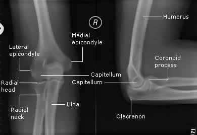
A review of the bony features of the elbow.
TRAUMATIC ELBOW INJURY
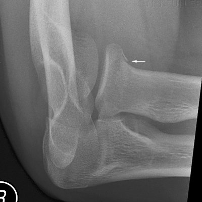
Most commonly from a FOOSH (fall on outstretched hand) injury, the radial head fracture is the winner of the most common injury in this joint. With the aforementioned mechanism and tenderness over the radial head (and pain with pronation/supination), have a low threshold to diagnose with occult fracture (even if one is not seen on plain films). As seen below, the presence of a posterior fat pad or sail sign indicates swelling (in this case bleeding) into the joint capsule and could indicate a radial head fracture and thus should be treated as such until proven otherwise. By way of review, a posterior fat pad seen on lateral films is never normal and an anterior fat pad can be normal but shouldn’t be a large triangle like a sail anteriorly. Little did I know, but you can also get a radial head-capitellum view plain x-ray in addition to standard views to pick up a few more radial head fractures not seen on initial films. Plain films are not perfect so splint all your patients with a strong likelihood of radial head fracture by history, exam, and/or x-rays in a long arm splint at 90 degrees flexion down to the wrist to prevent pronation and supination. These patients should be seen by ortho urgently as they might need surgery.
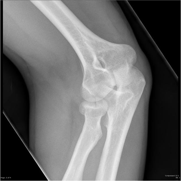
Radial head capitellum view, aka 45 degree radial head view from http://www.wikiradiography.net/page/Imaging+Radial+Head+Fractures
Other traumatic injuries include olecranon fracture and elbow dislocation. Both are easily seen on x-rays. The former occurs usually with a fall directly onto a flexed elbow (the x-ray finding and exam is obvious) and the latter takes substantial force to dislocate this relatively stable joint (and it takes a fair amount of force to reduce as well).
I have found the technique to the right to be the most effective, but if less muscle mass the other techniques would work.
What to document:
R/L handedness
Mechanism of injury
Radial pulse and capillary refill distally
Neuro exam of the wrist and hand
Bony tenderness or deformity of the medial/lateral epicondyl, olecranon, radial head and down the radius and ulna to the wrist.
What to do:
Splint fractures and suspected fractures at 90 degrees flexion. Per usual check and document neurovasc status after application
Provide pain relief up to procedural sedation for elbow reduction
What not to miss:
Test the radial, ulnar, and median nerves separately (see above) as they all cross the joint and could be injured in trauma
Feel radial pulse distally and consider an Allen test for possible ulnar artery injury.
Always think potential compartment syndrome and look for signs as well as warm patient of symptoms and when to return for a recheck.
Example PE for elbow trauma:
Musculoskeletal: The *** elbow has *** deformity. There is bony tenderness over the ***. Flexion and extension of the elbow is limited to ***. Strong wrist flexion and extension. No swelling of the olecranon bursa. The shoulder is not swollen or deformed on inspection and range of motion is full. There is no bony tenderness along the middle and distal radius or ulna. Sensation intact over the lateral deltoid and down to the fingertips including the palm and dorsum of the hand. Positive radial pulse. Normal range of motion and 5+ strength to flexion and extension of the wrist and fingers including finger adduction and abduction with good opposition of the thumb. Fingers are warm and well perfused and sensation intact.


